

Next: About this document
Up: Groping for Quantitative
Previous: Preliminary Results and Conclusions
References
- 1
-
Aubele, M., Zitzelsberger, H., Szücs, S., Werner, M., Braselmann, H., Hutzler, P.,
Rodenacker, K., Lehmann, L., Minkus, G., Höfler, H.:
Comparative FISH analysis of numerical chromosome 7 abnormalities in 5 m and
15 m paraffin-embedded tissue sections from prostatic carcinoma.
Histochem Cell Biol 107 (1997) 121-126
- 2
-
Becker Jr., C.R.L., Mikel, U.V., Oliver, LTC W.R., Sesterhenn, I.A.:
Enumeration of interphase chromosomes:
Comparison of visual in situ hybridization and confocal
fluorescence in situ hybridization.
Anal Quant Cytol Histol 18 5 (1996) 405-409
- 3
-
Babu, V.R., Miles, B.J., Cerny, J.C., Weiss, L., van Dyke, D.L.:
Cytogenetic study of four cancers of prostate.
Cancer Genet. Cytogenet. 48 (1990) 83-87
- 4
-
Irinopoulou, T., Vassy, J., Beil, M., Nicolopoulou, P., Encaoua, D.,
Rigaut, J.P.:
Three-dimensional DNA image cytometry by confocal scanning laser microscopy
in thick tissue blocks of prostatic lesions.
Cytometry 27 (1997) 99-105
- 5
-
Johnson, G.D., de C Nogueira Araujo, G.M.:
A simple method of reducing the fading of immunofluorescence during microscopy.
Immunol Methods 43 (1981) 349-350
- 6
-
Kriete, A. Ed.:
Visualization in biomedical microscopies -
3-d imaging and computer applications.
VCH, Weinheim (1992)
- 7
-
McNeal, J.E.:
Cancer volume and site of origin of adenocarcinoma of the prostate:
relationship to local and distant spread.
Hum. Pathol. 23 (1992) 258-266
- 8
-
Micale, M.A., Mohamed, A., Sakr, W., Powell, I.J., Wolman, S.R.:
Cytogenetics of primary adenocarcinoma.
Cancer Genet Cytogenet 61 (1992) 165-173
- 9
-
Pinkel, D., Straume, T., Gray, J.W.:
Cytogenetic analysis using quantitative high-sensitivity fluorescence hybridization.
Proc Natl Acad Sci USA 83 (1986) 2934-2938
- 10
-
Preston Jr., K. and Siderits, R.:
New technologies for 3-d data analysis in histopathology.
Anal Quant Cytol Histol. 14 5 (1992) 398-406
- 11
-
Scardino, P.T., Weaver, R., Hudson, M.A.:
Early detection of prostate cancer.
Hum. Pathol. 23 (1992) 211-222
- 12
-
Serra, J.:
Image analysis and mathematical morphology.
Academic Press, London (1982)
- 13
-
Sheppard, C.J.R and Gu, M.:
3-d transfer functions in confocal scanning microscopy.
In: [6] 251-281
- 14
-
Tekola, P., Baak, J.P.A., Beliën, J.A.M., Brugghe, J.:
Highly sensitive, specific, and stable new fluorescent DNA stains for
confocal laser microscopy and image processing of normal paraffin sections.
Cytometry 17 (1994) 191-195
- 15
-
Tekola, P., Baak, J.P.A., van Ginkel, H.H.A.M., Beliën, J.A.M., van
Diest, P.J., Broeckert, M.A.M., Schuurmans, L.T.:
Three-dimensional confocal laser scanning DNA ploidy cytometry in thick
histological sections.
J. Pathol. 180 (1996) 214-222
- 16
-
Vrolijk, H., Sloos, W.C.R., van de Rijke, F.M., Mesker, W.E., Netten, H.,
Young, I.T., Raap, A.K., Tanke, H.J.:
Automation of spot counting in interphase cytogenetics using brightfield
microscopy.
Cytometry 24 (1996) 158-166
- 17
-
Zhu, Q., Tekola, P., Baak, J.P.A., Beliën, J.A.M.:
Measurement by confocal laser scanning microscopy of the volume of
epidermal nuclei in thick skin sections.
Anal. Quant. Cytol. Histol. 16 2 (1994) 145-152
- 18
-
Zitzelsberger, H., Szücs, S., Weier, H.-U., Lehmann, L.,
Braselmann, H., Enders, S., Schilling, A., Breul, J.,
Höfler, H., Bauchinger, M.:
Numerical abnormalities of chromosome 7 in human prostate cancer
detected by fluorescence in situ hybridization (FISH) on
paraffin-embedded tissue sections with centromere-specific DNA probes.
J. Pathol. 172 (1994) 325-335

Figure 1:
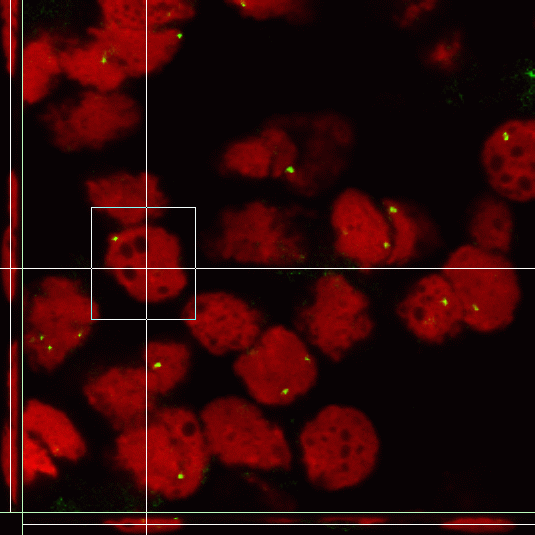
Graphical user interface for 3-D image display

Figure 2:
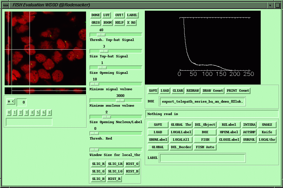
Functional tablet for user interaction,
a) Segmentation and semi-automatic count and journaling
b) Display, visual count and journaling

Figure 3:
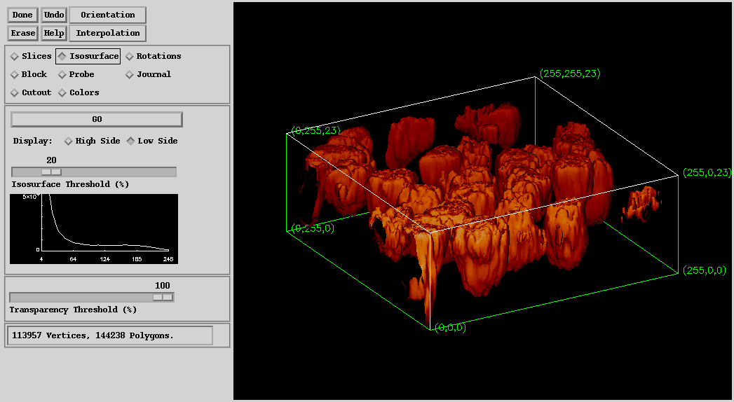
Graphical user interface for volume display by rendering and slicing

Figure 4:
Graphical user interface for volume display by rendering and slicing

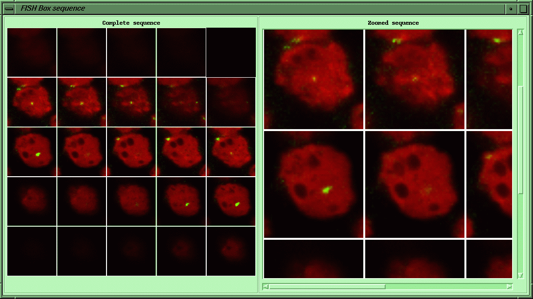
Figure 5: Gallery of a selected box

Figure 6:
Result display with
a) marked objects and
b) FISH signal distribution}

Figure 7:
Labelled section,
a) globally segmented,
b) locally improved by cutting touching nuclei,
c) cleaned by deletion
rodena_@_gsf.de
