After selection of the cell nucleus and its digitisation with a CCD-camera, the cell nucleus is segmented in the image and (at least during the development step) examined with a multitude of analysis procedures. Each procedure delivers quantitative features. These features are then correlated with external criteria such as survival time or diagnosis. The features with highest correlation or most discriminatory power are selected. This procedure is called "supervised learning". In later routine work the calculation of the chosen features only is necessary.

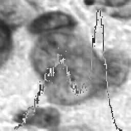
Section of a nucleus in transmission with frequency distribution
Which features are finally calculated ?
For the training phase extinction features like total sum (IOD = integrated optical density), mean value, standard deviation, entropy etc. from different areas of the cell nucleus are calculated. Extinction is physically related with mass of stain and hence its distribution reflects the arrangement of stain. E.g. with Feulgen stain the amount of DNA in the nucleus can be measured. The selection of different areas follow the procedures of visual observation of the pathologists to

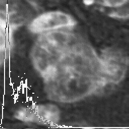
Nucleus in extinction with frequency distribution

Mask of nucleus with surrounding box and principal axies
The features mentioned describe in a statiscial manner the shape of frequency distributions. This is the reason that in the illustrations frequency distributions are mostly outlined.
In addition different procedures derived from pattern recognition for texture analysis are used. For a reproducible generation of border, eu- and hetero-chromatin areas, methods from the field of mathematical morphology are applied. The evaluation of images and their representation are carried out completely with IDL including several user written C routines.

Eu- and hetero chromatin particles with extinction frequency distribution

Flat texture image of the nucleus after 2D median filtering with frequency distribution