Institute of Computational Biology |

|
Institute of Computational Biology |

|
 Idl PROGRAMS for evaluation of cytometrical projects
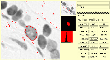
A cytometrical project consists of specimens (German Praeparat) and a parameter set for handeling the project. Each specimen consists of objects, in cytometry typically images of cells, eventually the mask images for each object and background images (German Weissbild). Objects are either digital images in our private format called DTP-file or tif-images. In DTP-file format the object image can contain in the least significant bit the mask for the region of interest. In scene mode this can be a set of regions. Tif-images need for this purpose a separate DTP-file containing the mask(s).
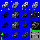
The evaluation of a project consists of the processing steps Image gathering (definition of objects), Image segmentation (definition of regions of interest), Feature evaluation (calculation of morphological, densitometrical, textural and achitectural features) and Statistical data evaluation (correlation of external project, specimen and object data with internal ones, supervised classification, unsuperwised classification, testing)
 PICKPROJECT
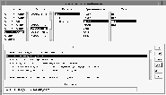
Graphical user interface (GUI) for general users for selection of projects, specimens and objects. The following programs can be activated from the GUI. References point to short descriptions.
 SCHWELLE_K

 PRAEPARAT_ZEIGEN

 PRAEPARAT_RECHNEN
Back to Image Cytometry home page

![]()
Last modified: 12.10.2014